
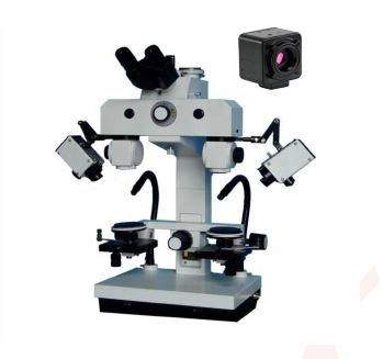
Steindorff Stereo Forensic Comparison Stereo Microscope with 5.0 Megapixels Camera,
Order Code: 23247121.5
Category: General Lab Equipment III
Technical Specifications:- Head - Comparison Bridge : 30° Inclined Trinocular Head with Two Photo/Video Ports. One port for still photography (use trinocular lever to divert light). One port for live video (always has light going to t...
SPECIFICATION
Technical Specifications:-
- Head - Comparison Bridge : 30° Inclined Trinocular Head with Two Photo/Video Ports. One port for still photography (use trinocular lever to divert light). One port for live video (always has light going to this port).
- Adjusts to the Distance Between your Eyes: 55 to 75mm Interpupillary Distance.
- Diopter Adjustment on Both Oculars to Correct for Your Specific Vision Needs.
- Adjustment screws on comparison bridge for adjusting the separation line width and separation line shape (if thicker on one end) on the split image view.
- Eyepieces and Magnification : Large Range of Magnifications from 4x up to 115x.
- Two Eyepiece Sets Included: 10x and 20x.
- Five Built-In Objectives on Rotary Selector Switchs: 0.8x, 1x, 2x, 3x, and 4.8x.
- Microscope Bridge Body Adds to Magnification by Factor of 1.2x.
- 10x Eyepiece Set Magnifications: 10x, 12x, 24x, 36x, 58x.
- 20x Eyepiece Set Magnifications: 19x, 24x, 48x, 72x, 115x.
- Install the Included 0.4x Bottom Screw-On Reduction Lens to Decrease Magnification! Use this Lens to Obtain Another Set of Magnifications for Each Eyepiece Set.
- Lowest Possible Magnification: Use the 10x Eyepiece and Bottom Reduction Lens: (0.8x Objective) (10x Eyepiece) (1.2 Bridge Factor) (0.4x Bottom Lens) = 3.8x Final Magnification. (Note: working distance will be long so some specimens with much height may not be focusable)
- Largest Possible Magnification: Use the 20x Eyepiece and Largest Power Objective:(4.8x Objective) (20x Eyepiece) (1.2 Bridge Factor) = 115x Final Magnification.
- Working Distance Approximately 105mm. Bottom lens 0.4x increases working distance.
- Illumination : Includes Two Illumination Methods.
- Light Source A: Variable Intensity 12V 50W Halogen Light Source in Box Enclosure.
- One halogen light mounted to each side, with a segmented arm position light.
- Light source has filter holder for accepting the included polarizing filters or colored filters.
- Light Source B: Variable Intensity Fiber Optic Light Source provides Incredible Illumination with Two Separate 12V 50W (100W total illumination power) lamps in the base light housing.
- Fiber Optic Cables come from base light housing and provide a Cool illumination on the mounting stage.
- Coaxial Illumination Feature: For illumination of the inside of a shell cartridge, inside of a hole, and some smooth surfaces, the coaxial illumination attachment is best. These attachments screw to the bottom of the objective lens and have openings for the fiber optic tips. The mirror the light downward, parallel to the optical light path.
- Transmitted Light Feature: Includes a stage attachment to provide light from the bottom, transmitting through the specimen. Useful for examining paper items, film negatives, money currency, postage stamps, fingerprints, etc. Use the side mounted box light source to shine on the transmitted light attachment's mirror to direct light under the specimen.
- Stage : Two Fully Rotatable Mechanical X-Y-Z Movable Stages, 65mm Diameter with Graduation Marks every 2° for the Full 360°.
- X-Y Stage Movement Knobs - Range of Movement: 51mm (X-Direction) x 51mm (YDirection) x 54mm (Z-Direction).
- Rack and Pinion Steel Gears with Knobs for Both X and Y Movements.
- Ball Socketed Inclinable Stages: Ball Socket Mounted Stages Provides Capability to Incline.
- Vice Type Mechanical Holder Attachments: Great for Holding Objects of Various Diameters, such as Bullets, Coins, and Industrial/Engineering Materials. Object can be Rotated as well as Inclined for Ease of Examining the Surfaces. Sits Directly on Stage. Two Included, One for Each Stage.
- Filters - Light Collectors : Side light sources have built-in filter holders.
- Color Filters Included: Red and Green.
- Polarizing Lens Filters: Polarized lens for mounting in the side lights with acorresponding polarized filter (called the analyzer) that mounts under the objective lens. This gives ability for doing cross polarization and is effective in increasing visual contrast on bright, shiny specimens, making it easier to see scratches and markings.
- Focusing : Focusing Knobs on Both Sides of Microscope.
- Stage Bridge Frame Focusing Knob Moves Both Stages Up/Down Simultaneously.
- Stage Bridge Frame Also has Coaxial Knob for Moving Both Stages Left/Right Simultaneously.
- Focusing Adjustment Travel Range for Bridge: 53mm. (Stage Bridge Frame Movement Distance Up/Down).
- Focusing Adjustment Travel Range for Stage: 53mm. (Individual Stage Movement Distance Up/Down).
- Video Port and Camera Details : High Resolution 5.0 MegaPixel Digital Camera System.
- Complete Digital Microscopy Solution Included.
- Capture high resolution digital microscope images, 2592 x 1944 pixels.
- View and record full motion live video microscope images. Frame rate: 30fps@2592 x 1944
- Computer connected digital microscope camera connects via USB2.0. Includes USB cable and MS Windows software.
- Includes measuring capability with the software.
Enquiry Form
Related Product
Tesca specialize in doing turnkey projects that is fully operable when it is handed over to the project authority. Starting from inception to application training, Tesca provides the services as ONE source solution. Working side by side with government authorities and people across the World, we help countries to perform better. We support countries grow their economies, strengthen their education and health systems and improve financial management. We do this by providing consultancy & training in environment safety, education, health strengthening.
Category
Useful Links
Contact Us
International Sales:
 91-9829132777
91-9413330765
India Sales:
91-9588842361
2024 © All Rights Reserved.