
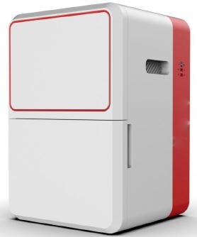
Stand-Alone Imaging System For Gel Documentation
Order Code: 25269410.42
Category: General Lab Equipment V
Stand-Alone Imaging System For Gel Documentation
SPECIFICATION
Stand-Alone Imaging System For Gel Documentation
- A gel documentation system is used in molecular biology labs to capture high-resolution images of stained nucleic acid or protein gels after electrophoresis. It consists of a camera, a light source (like a UV transilluminator), and a darkroom hood to visualize and analyze bands, providing data for record-keeping and research publications.
- Main Specifications - Versatile system to support wide range of applications like Fluorescence, Colorimetry /densitometry & Gel documentation, Suitable for DNA and RNA gels and fluorescence stain imaging
- Standalone system with large display and imaging
- Camera with resolution of at least 5 megapixels resolution with Passive cooling and Motorized zoom lens.
- White light and UV transilluminator
- The Gel Imaging System should offer precalibrated focus for any zoom setting or sample height.
- The Gel Placement door should be drawer type allowing access to Gels from either direction for facilitating easy/clutter free gel excision applications.
- A Slide-Out Transilluminator
- At least 4 positions filter wheel
- Compatible software for image acquisition, Molecular weight calculation, band quantification, colony counting, distance calculation, text annotation and image enhancement.
- System should have in-built touchscreen with Larger 12.6” display for image acquisition, Protein visualization and further quantification, documentation and publication should have software.
- System should have built-in computer-controlled, high-resolution scientific grade 16 bit CMOS camera, lens, lighting, darkroom and interchangeable UV and Blue light table options
- System should have USB 3.0 technology for fast image transfer
- System should have passive cooling (camera air circulation) for significant background noise reduction
- System should have dark room and EPI White LED along with UV light
- System should be made of Stainless Steel, Rust Free, covered with chemical-resistant Epoxy paint
- System should have UV Trans illuminator of dimension at least 26 x 21 cm FOV image area and Slide out Super Bright transilluminator to get background noise free images. . The Instrument should offer Trans-UV (B) and Epi White as Standard
- System should have multiuser Software for analysis with local area network (LAN) connectivity
- System should have remote access for image transfer and further analysis
- System should have Image Master Assistant - powerful tool to control sample image quality. A modification of the image display does not modify the Image Master data
- System should have One-touch fully automated image acquisition process
- System should have Auto-exposure, manual-exposure and serial modes - 3D and 3D-live image acquisition view mode
- System should have option for Image enhancement, annotation, illustration and comparison, Molecular weight, Dendrogram
- System should have option for UV to white light and UV to blue light conversion screen
- The Gel Imaging System should support the following dyes – SYBR Green, SYBR Safe, Ethidium Bromide, Stain Free Gel , Coommassie Blue, Zinc Stain, Flamingo, Oriole, Silver Stain, Coommassie Fluor Orange, Sypro ruby, Krypton & amp; Colorimetric Blots
- System should have option for Colorimetric stained protein gels, X-Ray film, autorads, SSCP gels, colony dish and flask imaging and other EPI white light applications
- System should have power requirements should be 220-240V
Specifications of the Image Analysis Software:-
- Automated lane and band identification, molecular weight or base pairs evaluation, band sizing, and quantitation based on a reference band or quantity standards
- Snapshot tool to copy images, lane profiles, and graphs
- Allow Publishing resolution (dpi) and publishing dimension to be specified with a one-click image export for publication. Provides functionality to produce image at user-defined dpi and dimension
- No requirement of license for registration. The full version software should be installable in large number of computers. Lifetime free upgrades of Software & Firmware should be available.
- Mac and PC compatible software
- 16-bit and 8-bit tiff images with a one-click export option
- Software should produce customizable reports with data organized as desired, including, Lane and band identification, molecular weight or base pair evaluation. Band sizing and quantification are based on a reference band or quantity standards.
- Software should offer live update of results with any change of analysis parameters.
- Local/Global background subtraction for individual bands
- Tools for compliance with U.S. FDA 21 CFR Part 11 regulations
Accessories
- Provided with compatible laptop.
- Provided with suitable UPS with half an hour backup
Enquiry Form
Related Product

Tesca specialize in doing turnkey projects that is fully operable when it is handed over to the project authority. Starting from inception to application training, Tesca provides the services as ONE source solution. Working side by side with government authorities and people across the World, we help countries to perform better. We support countries grow their economies, strengthen their education and health systems and improve financial management. We do this by providing consultancy & training in environment safety, education, health strengthening.
Category
Useful Links
Contact Us
International Sales:
 91-9829132777
91-9413330765
India Sales:
91-9588842361
2026 © All Rights Reserved.